US-based HR technology startup · HR technology & recruitment
OpenInterview
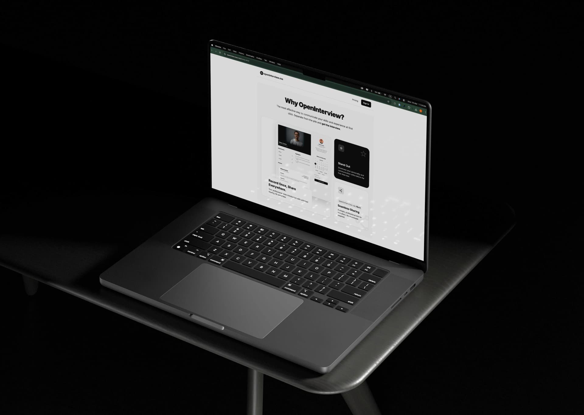
OpenInterview is a video interview platform that turns a candidate résumé into a recruiter ready profile with a video introduction availability details and one shareable link. Barakode built the product from résumé parsing and video processing to public profiles and Stripe subscriptions.
- Delivered
- 4 services
- Timeline
- August 2024 – November 2024
- React.js
- Node.js
- REST API
- AI résumé processing
- Cloud storage
Platform launched and live at openinterview.me, serving job seekers internationally.
View case study